OCT is normally used to obtain sub-surface images of biological tissue at about the same resolution as a low-power microscope and can instantly deliver cross-section images of layers of tissue without surgery or ionizing radiation. Images are based on light directly reflected from a sub-surface.
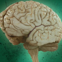
Widely used in clinical ophthalmology, OCT has recently been adapted for brain imaging in small animal models. Its application in neuroscience has been limited, however, because conventional OCT technology hasn’t been able to image more than 1 millimeter below the surface of biological tissue.
In the paper, Choi and Wang describe how a new technique called “swept-source OCT” (SS-OCT) powered by a vertical-cavity surface-emitting laser (VCSEL) increases signal sensitivity, extending the imaging depth range to more than 2 millimeters. That may make it possible to do things that have been barely attempted in the OCT community, such as noninvasive imaging of the mouse hippocampus or full-length imaging of a human eye from cornea to retina.
It could also allow researchers to monitor deeper morphological changes caused by diseases such as Alzheimer’s disease and dementia, and even to study the effects of aging on the brain.