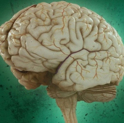
Visualizing human brain tissue in vibrant transparent colors: Neuroscientists from The University of Hong Kong (HKU) and Imperial College London have developed a new method called “OPTIClear” for 3D transparent color visualization (at the microscopic level) of complex human brain circuits.
To understand how the brain works, neuroscientists map how neurons (nerve cells) are wired to form circuits in both healthy and disease states. To do that, the scientists typically cut brain tissues into thin slices. Then they trace the entangled fibers across those slices — a complex, laborious process.
Making human tissues transparent. OPTIClear replaces that process by “clearing” (making tissues transparent) and using fluorescent staining to identify different types of neurons. In one study of more than 3,000 large neurons in the human basal forebrain, the researchers were able to reduce the time from about three weeks to five days to visualize neurons, glial cells, and blood vessels in exquisite 3D detail. Previous clearing methods (such as CLARITY) have been limited to rodent tissue.
Watching millions of brain cells in a moving animal for the first time:
It’s a neuroscientist’s dream: being able to track the millions of interactions among brain cells in animals that move about freely — allowing for studying brain disorders. Now a new invention, developed at The Rockefeller University and reported today, is expected to give researchers a dynamic tool to do that just that, eventually in humans.
The new tool can track neurons located at different depths within a volume of brain tissue in a freely moving rodent, or record the interplay among neurons when two animals meet and interact socially.
Microlens array for 3D recording. The technology consists of a tiny microscope attached to a mouse’s head, with a group of lenses called a “microlens array.” These lenses enable the microscope to capture images from multiple angles and depths on a sensor chip, producing a three-dimensional record of neurons blinking on and off as they communicate with each other through electrochemical impulses. (The mouse neurons are genetically modified to light up when they become activated.) A cable attached to the top of the microscope transmits the data for recording.
One challenge: Brain tissue is opaque, making light scatter, which makes it difficult to pinpoint the source of each neuronal light flash. The researchers’ solution: a new computer algorithm (program), known as SID, that extracts additional information from the scattered emission light.
Researchers at the David Geffen School of Medicine at UCLA can now peer deep inside a mouse’s brain to watch how star-shaped astrocytes (support glial cells in the brain) interact with synapses (the junctions between neurons) to signal each other and convey messages.
The method uses different colors of light that pass through a lens to magnify objects that are invisible to the naked eye. The viewable objects are now far smaller than those viewable by earlier techniques. That enables researchers to observe how brain damage alters the way astrocytes interact with neurons, and develop strategies to address these changes, for example.
Astrocytes are believed to play a key role in neurological disorders like Lou Gehrig’s, Alzheimer’s, and Huntington’s disease.