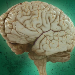
Two advances in detection technologies could soon up the odds of surviving cancers of the brain, head and neck. In one breakthrough, researchers have used an imaging technique that uses safe near-infrared light to differentiate between healthy brain tissue and that which is cancerous.
They hope to advance their work so that neurosurgeons can see 3-D color-coded maps of patient brains while they are performing surgery to more accurately remove tumors. In another, scientists were able to detect tumor DNA in the saliva and blood of people suffering from head and neck cancers. In diagnostic tests using both body fluids, they were able to successfully identify cancers in 96 percent of patients.
In the first research announcement, biomedical engineers and neuroscientists say they have used an imaging technique called optical coherence tomography to peer into brain tissue and see the difference between cancerous cells and normal. OCT has been used for years to identify disease in the retina, gastric tract, heart and breast.
The technique shines near-infrared light on the tissue and records differences in how different elements scatter the light waves. Johns Hopkins researchers refined the method so it can be used for brain surgery. They did it by relying on two characteristics of brain cancer cells: they form tumors that tend to be denser than healthy tissue and they don’t have the normal myelin sheaths that coat healthy cells. These facts mean that tumors scatter light differently than surrounding unaffected tissue, a property that can be detected by OCT.
The team developed algorithms that convert the signal differences picked up by the OCT sensor into a 3-D map of tissue color-coded red for cancerous tissue and green for healthy.
“We envision that the OCT would be aimed at the area being operated on, and the surgeon could look at a screen to get a continuously updated picture of where the cancer is, and isn’t,” said biomedical engineer Xingde Li, the coauthor of the study that appeared last week in the journal Science Translational Medicine.
They expect to begin clinical trials on the device this summer. They’ve already seen success in a proof of concept using human brain tissue removed during normal surgeries and performing their own surgeries on mice with brain tumors.