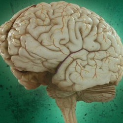
Specific areas of the brain are activated more strongly during lucid dreaming than in a normal dream, including these in the right hemisphere. Neuroscientists from the Max Planck Institutes of Psychiatry in Munich, Human Cognitive and Brain Sciences in Leipzig, and Charité in Berlin have identified a specific cortical network associated with self-awareness.
They used EEG and fMRI brain imaging to study “lucid dreamers,” who have access to their memories during dreaming and are aware of themselves, although remaining in a dream state and not waking up.
The researchers found neural activations in a specific network that is normally deactivated during REM sleep, comprising these areas:
Dorsolateral prefrontal cortex (associated with self-focused metacognitive evaluation)
Dorsolateral prefrontal cortex in combination with parietal lobules (may reflect working memory demands)
Bilateral frontopolar areas (related to the processing of internal states, e.g., the evaluation of one’s own thoughts and feelings)
Precuneus (implicated in self-referential processing, such as first-person perspective)
Bilateral cuneus and occipitotemporal cortices (active in conscious awareness in visual perception)
The study was limited to only four subjects who were highly trained lucid dreamers ”due to the rarity of lucid dreaming in untrained subjects, the researchers report. “Only one of them became lucid twice under concurrent EEG/fMRI conditions, rendering our data a case study.” The researchers also advised that part of the observed activation may have originated from the eye-signaling and hand-clenching task performed during the lucid-dreaming process.
