Surgeons in London have used lasers to diagnose abnormal tissue during an operation to remove a brain tumour for the first time in Europe. The non-invasive technique measures light reflected off tissue to determine whether it is cancerous or healthy. The patient, Reuben Hill, is recovering after the operation.
It is hoped the technique could make this kind of delicate surgery faster and more accurate. It has only been tried in Montreal, Canada, before now. Mr Hill, who is from Devon and studying for a PhD in physics at Imperial College London, works with the same laser technology as used in his operation.
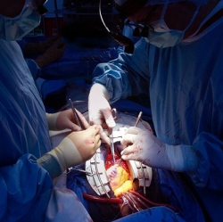
Before surgery, he told me: "My inner scientist is fascinated by what they are going to do. "Understanding the physics involved definitely makes it less frightening." During the operation, surgeons use a near-infrared laser probe, pointing the beam of light on to the exposed brain.
This causes molecules in the cells to vibrate. Fibre optics in the probe collect the scattered light that bounces off the tissue. This is analysed using Raman spectroscopy, which can measure the frequency of vibrations.
Healthy and abnormal tissue have a slightly different "signature". The whole process takes a couple of seconds, and is entirely non-invasive. In all cancer surgery, the aim is to remove all abnormal tissue while sparing healthy cells.
This is especially important with brain tumours, as removing healthy tissue can cause permanent damage to cognition, memory and speech. The Raman probe can tell surgeons whether to cut or spare tissue. At present, they rely on biopsies sent for analysis during the operation, which can take up to 40 minutes each.