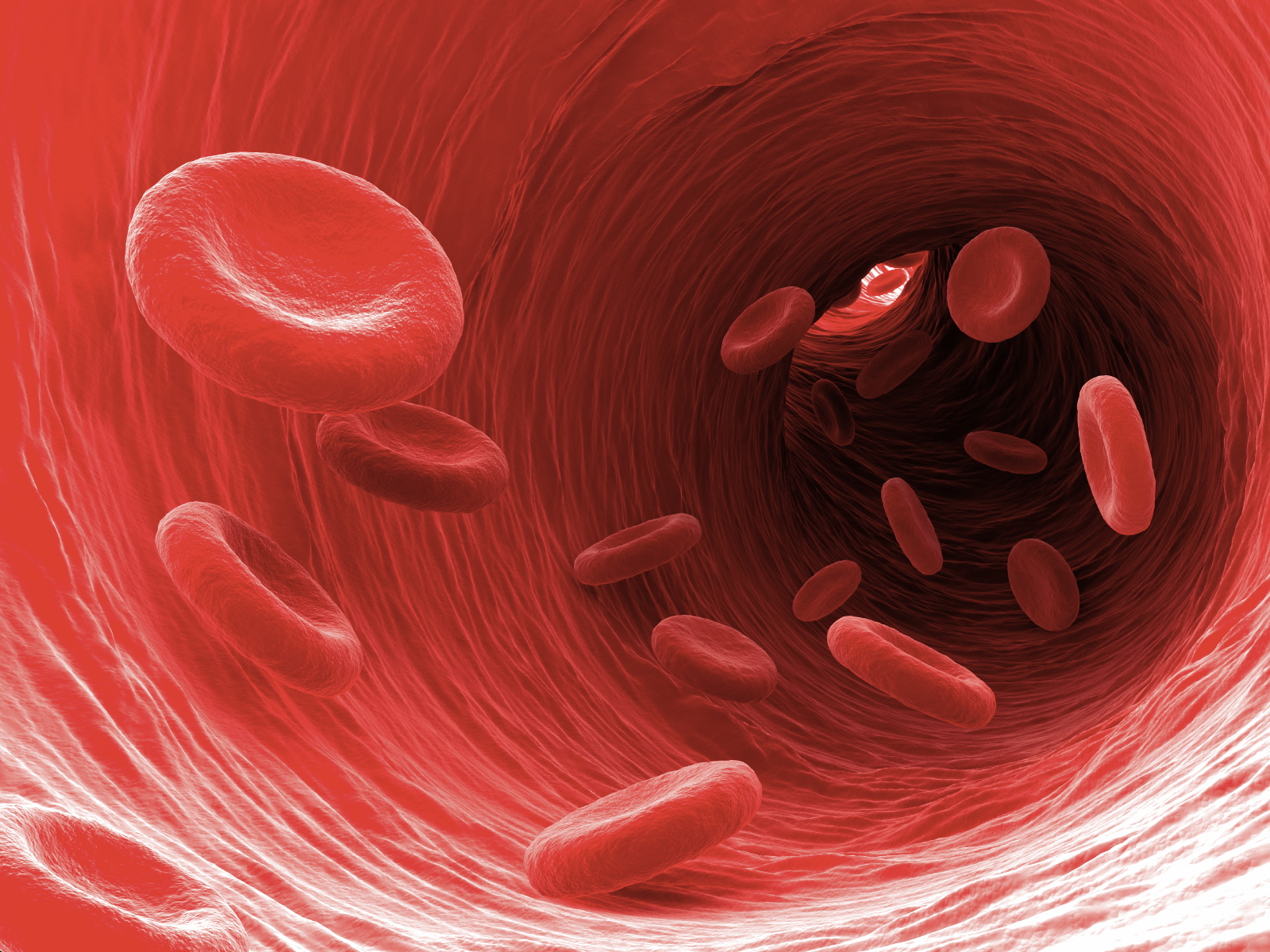
Reseachers have identified a biomarker for early detection of liver cancer. With current tests like biopsies, cancer in the liver is often detected in advanced stages, which can limit treatment options and lower overall survival rates. The discovery could have wide-ranging impacts, including more effective diagnosis and precision treatment, and less risk to patients.
“This is a game changer. It has the possibility to have many more applications, really for any type of cancer,” said Yang. “We are already applying it to 10 different types of cancer in the lab.”
They hope within 18 months to two years to conduct first clinical trials in patents.
The research builds on a 2019 study published in the journal Nature Communications which identified the first early detection of liver fibrosis using a collagen-targeted protein contrast agent.
Liver metastases often progress from primary cancers including uveal melanoma (UM), breast, and colon cancer. Molecular biomarker imaging is a new non-invasive approach for detecting early stage tumors. Here, we report the elevated expression of chemokine receptor 4 (CXCR4) in liver metastases in UM patients and metastatic UM mouse models, and development of a CXCR4-targeted MRI contrast agent, ProCA32.CXCR4, for sensitive MRI detection of UM liver metastases. CXCR4 molecular imaging capability in metastatic and intrahepatic xenotransplantation UM mouse models. ProCA32.CXCR4 enables detecting UM liver metastases as small as 0.1 mm3. Further development of the CXCR4-targeted imaging agent should have strong translation potential for early detection, surveillance, and treatment stratification of liver metastases patients
Uveal melanoma (UM) is the most common primary intraocular malignancy in adults. Approximately 50% of UM patients will develop metastases. About 93% of UM metastases occur in the liver, which results in death in almost all cases due to the lack of effective treatments.
The liver is a common site for cancer metastases. UM almost exclusively metastasizes to the liver. The mechanism of the liver-specific metastases is not well understood. One of the hypotheses in the field postulates that tumor cells that overly express CXCR4 hijack the CXCR4/CXCL12 axis during the metastatic process and spread to the liver. This hypothesis is based on the findings that the liver microenvironment in UM is rich in multiple chemoattractants including CXCL12, the natural ligand of CXCR4, and CXCR4 was found to be overexpressed on UM cells in several UM cell line studies. CXCR4 is proposed to be a prognostic marker in multiple malignancies, including acute myelogenous leukemia, breast cancers, colorectal cancers, and cutaneous melanoma. Thus, development of imaging agents for CXCR4 may be used as a diagnostic biomarker in cancer and potentially as a prognostic factor.
In this investigation, they validated the diagnostic value of CXCR4 as an imaging biomarker in UM by demonstrating elevated CXCR4 expression in three different biological systems: UM patient liver metastases, UM cell lines, and an in vivo UM murine model.