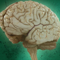
If research on the brain is to come close to meeting the hopes many researchers and government bodies have for it, one thing is going to have to change: Brain scanning will have to become a process that can be done in less restrictive situations and without the health risks that make some scanning methods an option of last resort.
Diffuse optical technology, or DOT, is closing in on the accuracy of the industry standard: functional MRI. Though the fMRI is accurate, it requires the patient to be immobilized in the machine, restricting how useful it is for understanding how the brain responds to stimuli.
In a study recently published in Nature Photonics, researchers debuted a new DOT, or diffuse optical tomography, instrument that could see two-thirds of the head and depict brain processes that take place in multiple regions and brain networks, such as those involved in language. The researchers, led by Joseph Culver, a radiologist at Washington University of St. Louis, compared the maps of activity created by DOT and fMRI scans and found a 75 percent overlap.
DOT itself isn’t new. For about a decade, doctors and researchers have looked to it as a more portable and safer alternative to fMRI scans. MRI technology uses magnets which interfere with the functioning of medical devices including pacemakers and cochlear implants. PET scans, another common method of coaxing a brain to do the equivalent of say ah, expose patients to significant levels of radiation.
But because DOT uses light, rather than the MRI’s magnets, to observe increased blood flow to active neural areas, getting a full picture of what goes on in the entire brain has been the main challenge. (Here’s a useful illustration of how DOT works.) The technology only illuminates what goes on in the outermost centimeter of brain tissue, but that area includes some pretty compelling functions like memory and language.
“When the neuronal activity of a region in the brain increases, highly oxygenated blood flows to the parts of the brain doing more work, and we can detect that. It’s roughly akin to spotting the rush of blood to someone’s cheeks when they blush,” Culver said in a news release.
