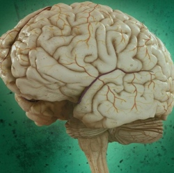
A new ultrasound device, used in conjunction with MRI (magnetic resonance imaging), allows neurosurgeons to precisely burn out small pieces of malfunctioning brain tissue without cutting the skin or opening the skull. A preliminary study involving nine patients with chronic pain shows that the technology can be used safely in humans.
The researchers now aim to test it in patients with other disorders, such as Parkinson’s disease.
“The groundbreaking finding here is that you can make lesions deep in the brain, through the intact skull and skin, with extreme precision and accuracy and safety,” says Neal Kassell, a neurosurgeon at the University of Virginia. Kassell, who was not directly involved in the study, is chairman of the Focused Ultrasound Surgery Foundation, a nonprofit based in Charlottesville, VA, that was founded to develop new applications for focused ultrasound.
High-intensity focused ultrasound (HIFU) is different from the ultrasound used for diagnostic purposes, such as prenatal screening. Using a specialized device, high-intensity ultrasound beams are focused onto a small piece of diseased tissue, heating it up and destroying it. The technology is currently used to ablate uterine fibroids–small benign tumors in the uterus–and it’s in clinical testing for removing tumors from breast and other cancers. Now InSightec, an ultrasound technology company headquartered in Israel, has developed an experimental HIFU device designed to target the brain.
The major challenge in using ultrasound in the brain is figuring out how to focus the beams through the skull, which absorbs energy from the sound waves and distorts their path. The InSightec device consists of an array of more than 1,000 ultrasound transducers, each of which can be individually focused. “You take a CT scan of the patient’s head and tailor the acoustic beam to focus through the skull,” says Eyal Zadicario, head of InSightec’s neurology program. The device also has a built-in cooling system to prevent the skull from overheating.
The ultrasound beams are focused on a specific point in the brain–the exact location depends on the condition being treated–that absorbs the energy and converts it to heat. This raises the temperature to about 130 degrees Fahrenheit and kills the cells in a region approximately 10 cubic millimeters in volume. The entire system is integrated with a magnetic resonance scanner, which allows neurosurgeons to make sure they target the correct piece of brain tissue. “Thermal images acquired in real time during the treatment allow the surgeon to see where and to what extent the rise in temperature is achieved,” says Zadicario.
The Swiss study, published this month in the Annals of Neurology, tested the technology on nine patients with chronic debilitating pain that did not respond to medication. The traditional treatment for these patients is to use one of two methods to destroy a small part of the thalamus, a structure that relays messages between different brain areas. Surgeons either use radiofrequency ablation, in which an electrode is inserted into the brain through a hole in the skull, or they use focused radiosurgery, a noninvasive procedure in which a focused beam of ionizing radiation is delivered to the target tissue.
Zadicario says HIFU has advantages over radiosurgery because the effects of killing tissue with radiation can take weeks to months, whereas the thermal approach is immediate. Adds Kassell, “The precision and accuracy [are] considerably greater with ultrasound, and it should be in principle safer in the long run.”